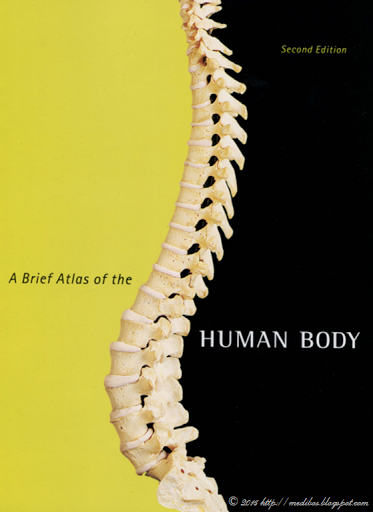
This full-color atlas is packaged with every new copy of the text, and includes 107 bone and 47 cadaver photographs with easy-to-read labels. This edition of the atlas contains a comprehensive histology photomicrograph section featuring over 50 slides of basic tissue and organ systems. Featuring photos taken by renowned biomedical photographer Ralph Hutchings, this high-quality photographic atlas makes an excellent resource for the classroom and laboratory, and is referenced in appropriate figure legends throughout the text.
English | ISBN: 032166261X | 2010 | 287 pages | PDF | 43 MB | 2nd


Không có nhận xét nào:
Đăng nhận xét