Latest News

Donate if you like this blog

Hiển thị các bài đăng có nhãn Radiology. Hiển thị tất cả bài đăng

Thứ Hai, 9 tháng 5, 2016

Merrill's Atlas of Radiographic Positioning and Procedures: 3-Volume Set 13th Edition


More than 400 projections make it easier to learn anatomy, properly position the patient, set exposures, and take high-quality radiographs! With Merrill's Atlas of Radiographic Positioning & Procedures, 13th Edition, you will develop the skills to produce clear radiographic images to help physicians make accurate diagnoses. It separates anatomy and positioning information by bone groups or organ systems - using full-color illustrations to show anatomical anatomy, and CT scans and MRI images to help you learn cross-section anatomy. Written by radiologic imaging experts Bruce Long, Jeannean Hall Rollins, and Barbara Smith, Merrill's Atlas is not just the gold standard in radiographic positioning references, and the most widely used, but also an excellent review in preparing for ARRT and certification exams!
UNIQUE! Collimation sizes and other key information are provided for each relevant projection.
Comprehensive, full-color coverage of anatomy and positioning makes Merrill's Atlas the most in-depth text and reference available for radiography students and practitioners.
Coverage of common and unique positioning procedures includes special chapters on trauma, surgical radiography, geriatrics/pediatrics, and bone densitometry, to help prepare you for the full scope of situations you will encounter.
Numerous CT and MRI images enhance your comprehension of cross-sectional anatomy and help you prepare for the Registry examination.
Bulleted lists provide clear instructions on how to correctly position the patient and body part when performing procedures.
Summary tables provide quick access to projection overviews, guides to anatomy, pathology tables for bone groups and body systems, and exposure technique charts.
Frequently performed projections are identified with a special icon to help you focus on what you need to know as an entry-level radiographer.
NEW! Coverage of the latest advances in digital imaging also includes more digital radiographs with greater contrast resolution of pertinent anatomy.
NEW positioning photos show current digital imaging equipment and technology.
UPDATED coverage addresses contrast arthrography procedures, trauma radiography practices, plus current patient preparation, contrast media used, and the influence of digital technologies.
UPDATED Pediatric Imaging chapter addresses care for the patient with autism, strategies for visit preparation, appropriate communication, and environmental considerations.
UPDATED Mammography chapter reflects the evolution to digital mammography, as well as innovations in breast biopsy procedures.
UPDATED Geriatric Radiography chapter describes how to care for the patient with Alzheimer’s Disease and other related conditions.
*****************
English | ISBN: 0323263410 | 2015 | 1771 pages | PDF | 178 Mb | 13th
****************
icon-download[7]

Thứ Tư, 27 tháng 4, 2016

Netter's Correlative Imaging - Abdominal and Pelvic Anatomy 1st Edition


2013 BMA Medical Book Awards Highly Commended in Radiology!
Visualize normal anatomy of the abdomen and pelvis like never before with Netter's Correlative Imaging: Abdominal and Pelvic Anatomy. This outstanding radiology reference from Dr. Drew A. Torigian and Dr. Mary Kitazono Hammell features beautiful and instructive Netter paintings and illustrated cross-sections created in the Netter style, presented side by side with high-quality patient MR images to help you envision and review both gastrointestinal and genitourinary anatomy section by section.
View organs, vessels, and peritoneal anatomy through MR, MRA, MRV, and MRCP imaging in a variety of planes, complemented with a detailed illustration of each slice done in the instructional and aesthetic Netter style.
Find anatomical landmarks quickly and easily through comprehensive labeling and concise text highlighting key points related to the illustration and image pairings.
Correlate patient data to idealized normal anatomy, always in the same view with the same labeling system.
Access NetterReference.com where you can quickly and simultaneously scroll through the book’s images and illustrations.
*****************
English | ISBN: 1437736548 | 2012 | 518 pages | PDF | 180 Mb | 1st
****************
icon-download[7]

Thứ Tư, 30 tháng 3, 2016

Spine Imaging A Case-Based Guide to Imaging and Management 1st Edition

Spine Imaging uses a classic, case approach to provide multi-modality imaging studies and management guidance of spine pathologies for the consulting radiologist as well as the neurologist, orthopedist, or emergency clinician diagnosing and managing spinal patients. Eighty cases, featuring over 650 images, are organized into sections by pathologic process and include a clinical description followed by discussion on radiologic findings, clinical management, and a summary of key points. An additional section on imaging signs closes the volume. Cases address which image findings are clinically significant and which are not, how to understand the severity of an injury, and when additional advanced imaging is needed, making Spine Imaging an essential resource for managing these complex pathologies.
*****************
English | ISBN: 019939394X | 2016 | 377 pages | PDF | 68.3 Mb | 1st
****************
icon-download[7]

Thứ Năm, 10 tháng 3, 2016

Radiologic-Pathologic Correlations from Head to Toe


We are proud to offer this unique textbook,the first in Radiology, The International Congress of Radiology, its class presenting state-of-the-art radiologic images of The Radiological Society of North America Assembly, the entire body correlated with underlying pathology. among others made us believe that radiologists,as well This book is based on a categorical course presented for as pathologists and other specialists,would enjoy hav- 3 years at recent European Congresses of Radiology and ing in a single source the best of class in this educati- covers from head to toe,key aspects of human disease al method. This book is indeed a global effort since it - lending themselves well to the correlation of modern sembles the leading authorities in all major organ imaging techniques with microscopic and gross pathol- systems who use radiologic-pathologic correlation as a ogy. Obviously, the most suitable candidate for radio- research and teaching method. Many of the chapter - logic-pathologic correlation is the study of neoplasms, thors have dedicated the bulk of their professional life to since they are resected ideally in toto and therefore offer educating radiologists throughout the world using this excellent gross-imaging comparisons. Although there is method. There are experts from 12 European countries a wealth of educational material based on radiolog- and the USA.

*****************
English | ISBN: 3540043950 | 2005 | 783 pages | PDF | 75.4 Mb | 2005Ed
****************
icon-download[7]

Chủ Nhật, 27 tháng 12, 2015

Clark's Positioning in Radiography 12th Edition

First published in 1939, this is the definitive text on patient positioning for the diagnostic radiography student and practitioner. The experienced author team appreciates that there is no substitute for a good understanding of basic skills in patient positioning and an accurate knowledge of anatomy to ensure good radiographic practice.
This 12th edition retains the book’s pre-eminence in the field, with hundreds of positioning photographs and explanatory line diagrams, a clearly defined and easy-to-follow structure, and international applicability.
The book presents the essentials of radiographic techniques in a practical way, avoiding unnecessary technical complexity and ensuring that the student and practitioner can find quickly the information that they require regarding particular positions. All the standard positioning is included, accompanied by supplementary positions where relevant and illustrations of pathology where appropriate. Common errors in positioning are also discussed.
            *****************
            English | ISBN: 0340763906 | 2005 | 532 pages | PDF | 28.4 Mb | 12th
            ****************
            icon-download[7]

            Interventional Radiology Cases - Cases in Radiology 1st Edition

            In 104 cases featuring over 500, high-quality images, Interventional Radiology Cases is a thorough and accessible review of the interventional procedures that radiology residents are expected to be familiar with upon completion of residency and general radiologists need to know for recertification examinations. The cases present both benign and malignant conditions and all pertinent imaging modalities incorporated including: CT, MR, PET, fluoroscopy, and ultrasound. Part of the Cases in Radiology series, this book follows the easy-to-use format of question and answer in which the patient history is provided on the first page of the case, and radiologic findings, differential diagnosis, teaching points, next steps in management, and suggestions for furthering reading are revealed on the following page.
                      *****************
                      English | ISBN: 0199331278 | 2015 | 337 pages | PDF | 60.3 Mb | 1st
                      ****************
                      icon-download[7]

                      Thứ Tư, 16 tháng 12, 2015

                      Gastrointestinal Imaging 1st Edition

                      Gastrointestinal Imaging presents a comprehensive review of gastrointestinal pathologies commonly encountered by practicing radiologists and residents in training. Chapters are organized by organ system and include the Pharynx and Esophagus, Stomach, Small Bowel, Appendix, Colon, Anorectum, Liver, Gallbladder, Bile Ducts, Pancreas, Spleen, Peritoneum, Mesentery, and Abdominal Wall, and a chapter on multisystem disorders. Part of the Rotations in Radiology series, this book offers a guided approach to imaging diagnosis with examples of all imaging modalities complimented by the basics of interpretation and technique and the nuances necessary to arrive at the best diagnosis. Each pathology is covered with a targeted discussion that reviews the definition, clinical features, anatomy and physiology, imaging techniques, differential diagnosis, clinical issues, key points, and further reading. This organization is ideal for trainees' use during specific rotations and for exam review, or as a quick refresher for the established gastrointestinal imager.
                          *****************
                          English | ISBN: 0199759421 | 2015 | 761 pages | PDF | 38.5 Mb | 1st 
                          ****************
                          icon-download[7]

                          Thứ Năm, 29 tháng 10, 2015

                          Chest Radiology 1st Edition

                          Chest radiology is the most commonly-used investigation method for pulmonary diseases. Accurate interpretation by radiologists is essential, in order to diagnose and manage disorders effectively. This atlas is a concise guide to chest radiology for residents and clinicians. Beginning with an introduction to anatomy, the book presents state of the art chest X-Ray, CT, MRI and PET scan images for numerous medical conditions. The book offers a clear understanding of how to recognise and interpret basic radiological signs, pathologies and patterns for differential diagnosis. A photo CD ROM is included to enhance the numerous well-illustrated X-Rays, CT and MRI scans in the atlas, making it an invaluable, hands-on reference for the evaluation of chest images. Key points * Concise guide to chest radiology for residents and clinicians * Presents numerous chest X-Rays, CT, MRI and PET scan images for chest diseases and disorders * Offers clear understanding of radiological signs, pathologies and patterns to assist differential diagnosis * Includes photo CD ROM

                        • English | ISBN: 9350904632 | 2013 | 226 pages | PDF | 25.2 Mb | 1st
                          Download this book here
                        • Thứ Năm, 22 tháng 10, 2015

                          Pearls and Pitfalls in Emergency Radiology: Variants and Other Difficult Diagnoses 1st Edition


                          Rapid recognition of life-threatening illnesses and injuries expedites appropriate management and improves clinical outcomes. False-positive interpretations in radiology have been identified as a significant cause of error, leading to unnecessary investigation and treatment, increased healthcare costs, and delays in appropriate management. Moreover, it is important that radiologists do not miss important subtle diagnoses that need urgent intervention. Pearls and Pitfalls in Emergency Radiology provides an outline of common imaging artefacts, anatomic variants and critical diagnoses that the radiologist must master in order to guide appropriate care and avoid malpractice lawsuits. One hundred selected cases - illustrated with several hundred images from MRI, MDCT, PET, ultrasound and radiographs - are presented in a succinct and structured format, highlighting key pearls and potential diagnostic pitfalls. The text focuses on emergent presentations of diseases in all body regions in both adults and children.
                        • English | ISBN: 110702191X | 2013 | 389 pages | PDF | 66.5 Mb | 1st
                          Download this book here
                        • Thứ Ba, 20 tháng 10, 2015

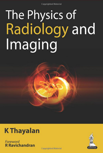
                          The Physics of Radiology and Imaging 1st Edition

                          This book explains the principles, instrumentation, function, application and limitations of all radiological techniques - radiography, fluoroscopy, mammography, computed tomography, ultrasound and magnetic resonance imaging. Beginning with an introduction to the fundamental concepts, the following chapters provide in depth coverage of each of the techniques from the perspective of a medical physicist. Presented in an easy to read format, this book is an invaluable reference for postgraduate students in medical physics and radiology and candidates training for FRCR exams. It includes nearly 280 images, illustrations and tables to enhance learning. Key points * Explains principles, instrumentation, function, application and limitations of all radiological techniques * Presented from perspective of medical physicists * Includes nearly 280 images, illustrations and tables * Highly useful for postgraduates in medical physics and radiology, and FRCR candidates

                          • English | ISBN: 9351521710 | 2014 | 426 pages | PDF | 17.5 Mb | 1st


                          Download this book here

                          Thứ Hai, 19 tháng 10, 2015

                          Pearls and Pitfalls in Pediatric Imaging Variants and Other Difficult Diagnoses 1st Edition


                          This collection of over 90 highly-illustrated case studies explores major and confusing problems in pediatric imaging. All relevant imaging modalities are covered, including ultrasound, conventional radiography, fluoroscopy, CT, MR, Nuclear and Molecular Imaging, and Interventional Radiology. The authors present a strategy for recognizing key information in order to reach an accurate diagnosis, and each case includes differential diagnoses and key teaching points, alerting the reader to common pitfalls in the interpretation of pediatric radiological images. This is a highly valuable resource for trainee pediatric radiologists, and general radiologists who encounter pediatric patients. It will particularly help people preparing for exams, including the core exam, the certifying exam or CAQ exams, as well as pediatric radiologists who want to refresh their knowledge on particular topics. It will also be of interest to pediatricians who wish to improve their diagnostic proficiency and understanding of imaging studies.
                          • English | ISBN: 1107017491 | 2014 | 270 pages | PDF | 15.8 Mb | 1st

                          Download this book here

                          Pearls and Pitfalls in Musculoskeletal Imaging 1st Edition

                          When a radiological image includes unfamiliar features, how do you decide whether it is normal variation or pathological abnormality? If you decide an abnormality is present, can you make a diagnosis from the image alone? Pearls and Pitfalls in Musculoskeletal Imaging differentiates less common findings or normal variant mimickers from the more common similar appearing diseases, helping you make a quick and accurate diagnosis. Musculoskeletal disorders of the shoulder, upper extremity, pelvis, and lower extremity are described in over 90 cases, highly illustrated with over 300 radiographic, CT, MRI and ultrasound images. Each case follows a standard format: imaging description, importance, typical clinical scenario, differential diagnosis and teaching point, enabling you to locate key information quickly. Pearls and Pitfalls in Musculoskeletal Imaging will help you spot artifacts, mimics and other unusual conditions, enabling you to avoid misdiagnosis and prevent mismanagement. An essential diagnostic tool for radiologists at every level. 

                          • English | ISBN: 0521196329 | 2013 | 270 pages | PDF | 15.8 Mb | 1st


                          Download this book here

                          Chủ Nhật, 18 tháng 10, 2015

                          Pearls and Pitfalls in Head and Neck and Neuroimaging: Variants and Other Difficult Diagnoses 1st Edition

                          Pearls and Pitfalls in Head and Neck and Neuroimaging illustrates and describes the imaging entities that can cause confusion and mismanagement in daily radiological practice. Frequent interpretation errors are covered in 105 cases that provide real-life clinical scenarios for focused and practical learning. The chosen cases represent a modern neuroradiology practice and cover the brain, spine and head and neck regions, including the full panoply of imaging modalities. The underlying reasons for common mistakes are analyzed and radiologic findings that help with the correct diagnosis are emphasized. A differential diagnosis is provided for each case with examples of alternative diagnoses, allowing readers to visually grasp the differentiating features. Pearls and Pitfalls in Head and Neck and Neuroimaging provides a valuable resource for general and sub-specialist radiologists needing to improve their diagnostic proficiency in adult and pediatric patients. This book also serves as a preparation resource for recertification exams in radiology.

                          • English | ISBN: 1107026644 | 2014 | 504 pages | PDF | 45.3 Mb | 1st


                          Download this book here

                          Thứ Tư, 14 tháng 10, 2015

                          Fundamentals of Skeletal Radiology 4th Edition

                          Fundamentals of Skeletal Radiology remains a perfect first book on musculoskeletal radiology and a terrific quick review of the subject. With its entertaining writing style and many new and improved imaging examples, turn to the "pink book" for an effective, concise, and enjoyable introduction to musculoskeletal imaging - just as tens of thousands of radiology students, residents, and clinicians have done with previous editions of this medical reference book.

                          "A clear, concise and quick reference, dipping into the pages is like slipping on a favourite pair of slippers - comforting and reassuring!" (Tracey Thorne, Specialist reporting radiographer, Airedale NHS Foundation Trust - Sept14) "Some may lament the cover colour and although the fourth edition ‘pink book’ is a more subtle cerise these days, it is still the go-to guide for skeletal radiology and the pearls that every reporter needs in order to build a firm foundation of MSK knowledge " Reviewed by: RAD Magazine  Date: Sept 2014

                          "It is an excellent book for all students of medical imaging and one that I recommend to all those who are developing an interest in skeletal imaging". Reviewed by: Stephen Boynes, University of Bradford  Date: 2014

                          • Visually grasp musculoskeletal imaging concepts and techniques through hundreds of high-quality digital radiographs, MRIs, bone scans, and CT images.
                          • Easily understand the basics of skeletal radiology from the author's succinct, highly accessible writing style that makes information straightforward for beginners.
                          • Quickly grasp the MSK radiology fundamentals you need to know through an easy-to-understand format and hundreds of radiographs and images.
                          • Discern subtleties and nuances by examining full-color imaging examples.
                          • Apply the latest knowledge and techniques in skeletal imaging. Extensive updates equip you with new technology and major advancements as well as an increased emphasis on MR imaging and enhanced coverage of knee imaging.
                          • Address radiation dosage concerns and apply new techniques aimed at early detection.
                          • English | ISBN: 1455751545 | 2013 | 248 pages | PDF | 107 Mb | 4h


                          Download this book here

                          Thứ Tư, 7 tháng 10, 2015

                          Clinical Radiology: The Essentials 4th Edition

                          Written in an engaging, easy-to-read style, Clinical Radiology covers the topics most often included in introductory radiology courses and emphasizes clinical problem solving. The text offers guidelines for selecting imaging studies in specific clinical situations and takes a systematic approach to imaging interpretation, presenting a review of normal anatomy, technical and pathologic considerations, and diagnostic advice.
                          The Fourth Edition includes:
                          -NEW! Full-color design and illustrations
                          -50 new images, updated to reflect the latest technology
                          -Expanded coverage of neurotoxicity and radiation exposure
                          -Additional "Diagnostic Pearls"  included in each chapter
                          -Bonus online material, including case studies, slides, and additional radiological images

                          • English | ISBN: 0729541479 | 2013 | 582 pages | PDF | 81.6 Mb | 4th


                          Download this book here

                          Thứ Ba, 22 tháng 9, 2015

                          Learning Radiology: Recognizing the Basics, 3rd Edition

                          A must-have for anyone who will be required to read and interpret common radiologic images, Learning Radiology: Recognizing the Basics is an image-filled, practical, and easy-to-read introduction to key imaging modalities. Skilled radiology teacher William Herring, MD, masterfully covers exactly what you need to know to effectively interpret medical images of all modalities. Learn the latest on ultrasound, MRI, CT, patient safety, dose reduction, radiation protection, and more, in a time-friendly format with brief, bulleted text and abundant high-quality images. Then ensure your mastery of the material with additional online content, bonus images, and self-assessment exercises at Student Consult.

                          • Identify a wide range of common and uncommon conditions based upon their imaging findings.
                          • Arrive at diagnoses by following a pattern recognition approach, and logically overcome difficult diagnostic challenges with the aid of decision trees.
                          • Quickly grasp the fundamentals you need to know through more than 700 images and an easy-to-use format and pedagogy, including: bolding of key points and icons designating special content; Diagnostic Pitfalls; Really, Really Important Points; Weblinks; and Take-Home Points.
                          • Gauge your mastery of the material and build confidence with extra images, bonus content, interactive self-assessment exercises, and USMLE-style Q&A that provide effective chapter review and quick practice for your exams.
                          • Apply the latest recommendations on patient safety, dose reduction and radiation protection
                          • Benefit from the extensive knowledge and experience of esteemed author Dr. William Herring-a skilled radiology teacher and the host of his own specialty website, www.learningradiology.com.
                          • Stay current in the latest advancements and developments with meticulous updates throughout including a new chapter on Pediatric Radiology as well as more than 60 new and updated photos, many highlighting newer imaging modalities.
                          • Maximize your learning experience with interactive Student Consult extras videos/images of 3D images, functional imaging examinations, dynamic studies, and additional assessments.
                          • Student Consult eBook version included with purchase. This enhanced eBook experience allows you to search all of the text, figures, references, and videos from the book on a variety of devices.

                        • English | ISBN: 0323328075 | 2015 | 451 pages | PDF | 71.9 Mb | 3rd
                          Download this book here
                        • Chủ Nhật, 20 tháng 9, 2015

                          Core Radiology: A Visual Approach to Diagnostic Imaging 1st Edition

                          Combining over 1200 clinical images, 300 color illustrations and concise, bulleted text, Core Radiology is a comprehensive, up-to-date resource for learning, reference and board review. The clearly-formatted design integrates the images and accompanying text, facilitating streamlined and efficient learning. 

                          All subjects covered by the American Board of Radiology Core Exam are included: 

                          • Breast imaging, including interventions and MRI
                          • Neuroimaging, including brain, head and neck, and spine 
                          • Musculoskeletal imaging, including knee and shoulder MRI
                          • Genitourinary imaging, including pelvic MRI
                          • Gastrointestinal imaging, including MRI and MRCP
                          • General, vascular, gynecological and obstetrical ultrasound
                          • Nuclear imaging, including PET-CT and nuclear cardiology
                          • Thoracic imaging
                          • Cardiovascular imaging, including cardiac CT and MRI
                          • Pediatric imaging
                          • Interventional radiology
                          • Radiological physics review, contrast media and reactions.
                          • Essential reading for radiology residents reviewing for boards, for practicing radiologists seeking a practical up-to-date guide to the field, and for motivated medical students.
                        • English | ISBN: 1609139712 | 2014 | 1417 pages | PDF | 56.2 Mb | 11th

                          Download this book here
                        • Thứ Ba, 15 tháng 9, 2015

                          Diagnostic Radiology: Gastrointestinal and Hepatobiliary Imaging 3rd

                          - Phenomenal technological advances and innovations in the last few years have prompted and necessitated a third series of server imaging courses and the reorgration of the accompanying book. - Ealier chapters have been thoroughly revised and updated inducing three new chapters covering the clinical aspects of liver cirrhosis, interventional treatment of liver tumors and percutaveous non-vascular GIT interventions. - The entire text has been divided into 2 partsgartiontertinal imaging and hepatobiolary and pancreatic imaging. Part I consists of Imaging techniques, acute abdomen, Infections, inflammation 2 neoplasm: part II contains Liver and bleary tract, pancreas, hepatic vascular diseases and Interventions. - The importance of plain radiography and barium studies has been described in depth. - Techniques of US CT and the potential role of MRI in evaluation of abdominal disorders have also been explicitly elaborated upon.

                        • English | ISBN: 8184484348 | 2009 | 452 pages | PDF | 17.5 Mb | 3rd
                          Download this book here
                        • Gastrointestinal Imaging: A Teaching File

                          A brand new resource in The Teaching Files series, Gastrointestinal Imaging: A Teaching File is an exceptional resource for radiology trainees and practicing radiologists who are interested in reviewing the basics of gastrointestinal imaging, including fluoroscopy, CT, and MR. Those studying for board and certifying examinations will also find this volume to be an excellent reference in helping them to review and prepare for these tests.
                          More than 300 cases, 100 in print and all 300 in an interactive ebook that is included with the print purchase, are portrayed through CT, MR, fluoroscopic, and ultrasound images.  This text also covers a wide variety of GI system disorders, including those that affect the solid abdominal organs, intestines, and peritoneal cavity. Each case includes images, along with descriptions of clinical history, findings, differential diagnosis and diagnosis, discussion, relevant questions with appropriate answers, key issues for the report, and important information for the clinician.
                          Features:

                          • More than 300 gastrointestinal, including 100 in print and all 300 in the interactive ebook help you make accurate and informed diagnoses.
                          • Fluoroscopy, CT, and MR images are featured.
                          • Case information includes images along with management discussion
                          • Cases are grouped into chapters based on modality to help trainees during modality-based rotations.
                            Now with the print edition, enjoy the bundled interactive eBook edition, offering tablet, smartphone, or online access to:
                          • Complete content with enhanced navigation
                          • A powerful search that pulls results from content in the book, your notes, and even the web
                          • Cross-linked pages, references, and more for easy navigation
                          • Highlighting tool for easier reference of key content throughout the text
                          • Ability to take and share notes with friends and colleagues
                          • Quick reference tabbing to save your favorite content for future use
                          • More than 400 Q&A
                        • English | ISBN: 1451173377 | 2014 | 207 pages | PDF | 10.3 Mb | 1st
                          Download this book here
                        • Chủ Nhật, 13 tháng 9, 2015

                          Radiology of Infectious Diseases: Volume 1 - 1st ed 2015 Edition

                          This book provides a comprehensive overview of diagnostic imaging in infectious diseases. It starts with a general review of infectious diseases, including their classification, characteristics and epidemiology. In separate chapters, the authors then introduce the radionuclide imaging of 50 kinds of infectious diseases. Volume 1 covers 21 viral infections. Volume 2 has 29 chapters discussing 24 bacterial infections and 5 parasitic infections. Each disease is clearly illustrated using cases combined with high-quality computed tomography (CT) and magnetic resonance imaging (MRI). The book provides a valuable reference source for radiologists and doctors working in the area of infectious diseases.

                        • English | ISBN: 9401798818 | 2015 | 390 pages | PDF | 46.3 Mb | 1st

                          Download this book here
                        • Translate

                          Total Pageviews