Thứ Hai, 13 tháng 6, 2016
Ultrasound in the Intensive Care Unit 2015 Edition

Thứ Bảy, 19 tháng 3, 2016
Differential Diagnosis in Ultrasound 2nd Edition

Thứ Ba, 8 tháng 3, 2016
Introduction to Musculoskeletal Ultrasound: Getting Started 1st Edition

Thứ Sáu, 4 tháng 3, 2016
Diagnostic Ultrasound: Musculoskeletal 1st Edition

- Readily accessible chapter layout with succinct, bulleted teaching points and almost 3,000 high-quality illustrative clinical cases and schematic designs.
- All-inclusive section on musculoskeletal ultrasound anatomy, as well as a comprehensive interventional section covering muskuloskeletal ultrasound.
- Approaches musculoskeletal ultrasound from two different viewpoints: that of a specific diagnosis (Dx section), followed by that of a specific ultrasound appearance (DDx section).
- Differential diagnosis section features supportive images and text outlining the key discriminatory features necessary in reaching the correct diagnosis.
- Provides a solid understanding of musculoskeletal ultrasound anatomy and pathology.
- Comes with Amirsys eBook Advantage™, an online eBook featuring expanded content, additional images, and fully searchable text.
Thứ Năm, 24 tháng 12, 2015
Pocket Protocols for Ultrasound Scanning, 2nd Edition

- Flip-card format with spiral binding allows the book to sit upright close to the console, so sonographer can easily flip pages while scanning to be sure that correct protocols are followed.
- Step-by-step instructions describe criteria for each ultrasound study, then provide examples of each scan that should be obtained to complete the study.
- Protocols follow guidelines provided by the American Institute of Ultrasound in Medicine (AIUM), the organization that sets sonography policies, practice standards, safety procedures, and performance guidelines.
- Detailed line drawings accompany most sonograms, clarifying the structures shown on the diagnostic image.
- Over 1,200 images and drawings are provided, including images with accompanying drawings for every AIUM-approved protocol - offering a visual step-by-step scanning approach to the performance of scans and image documentation for physician diagnostic interpretation.
- New images are added to complete the scanning protocols approved by the AIUM.
Thứ Tư, 28 tháng 10, 2015
Atlas of Pediatric Ultrasound 1st Edition

This atlas is a comprehensive guide to paediatric ultrasound. Each section discusses a different part of the body, with seven chapters dedicated to the abdomen. Each chapter begins with an explanation of technique, correct transducer use and offers guidance for achieving optimal imaging. More than 700 high quality ultrasound images are included, with detailed discussion on Doppler, and clearly marked to show the position and angle of the probe. Corresponding X-rays, CT scans and MRI images are also included to clarify and confirm the ultrasound pathologies.
Thứ Ba, 8 tháng 9, 2015
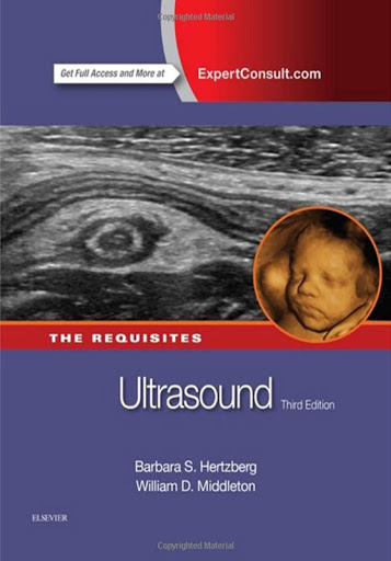
Ultrasound: The Requisites, 3e

This best-selling volume in The Requisites™ Series provides a comprehensive introduction to timely ultrasound concepts, ensuring quick access to all the essential tools for the effective practice of ultrasonography.
Comprehensive yet concise, Ultrasound covers everything from basic principles to advanced state-of-the-art techniques. This title perfectly fulfills the career-long learning, maintenance of competence, reference, and review needs of residents, fellows, and practicing physicians.
- Covers the spectrum of ultrasound use for general, vascular, obstetric, and gynecologic imaging.
- Fully illustrated design includes numerous side-by-side correlative images.
- Written at a level ideal for residents seeking an understanding of the basics, or for practitioners interested in lifelong learning and maintenance of competence.
- Extensive boxes and tables highlight differential diagnoses and summarize findings.
- "Key Features" boxes offer a review of key information at the end of each chapter.
- Explore extensively updated and expanded content on important topics such as practical physics and image optimization, the thyroid, salivary glands, bowel, musculoskeletal system, cervical nodal disease, ectopic pregnancy, early pregnancy failure, management of asymptomatic adnexal cysts, practice guidelines - and a new chapter on fetal chromosome abnormalities.
- Visualize the complete spectrum of diseases with many new and expanded figures of anatomy and pathology, additional correlative imaging, and new schematics demonstrating important concepts and findings.
- Further enhance your understanding with visual guidance from the accompanying electronic version, which features over 600 additional figures and more than 350 real-time ultrasound videos.
- Expert Consult eBook version included with purchase. The enhanced eBook experience allows you to view the additional images and video segments and access all of the text, figures, and suggested readings on a variety of devices.
Thứ Ba, 25 tháng 8, 2015
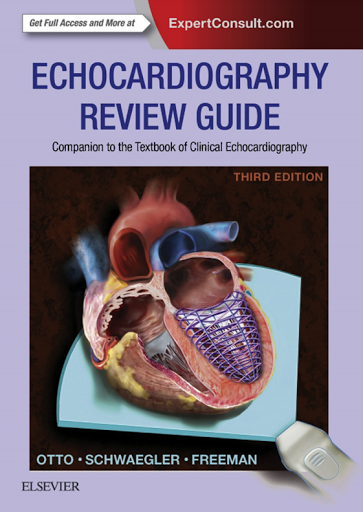
Echocardiography Review Guide: Companion to the Textbook of Clinical Echocardiography, 3e

This review companion to Dr. Catherine Otto’s Textbook of Clinical Echocardiography demonstrates how to record echos, avoid pitfalls, perform calculations and understand the fundamentals echocardiography for every type of cardiac problem. It teaches and tests in one convenient volume, with precise step-by-step instructions on using and interpreting echocardiography. It's a must-have for anyone new to the field or preparing for the echocardiography boards, the PTEeXAM, or the diagnostic cardiac sonographer’s exam.
- Enhance your calculation skills for all aspects of echocardiography.
- Multiple-choice questions in each chapter cover the latest information tested on exams.
- Features expert advice and easy-to-follow procedures on using and interpreting echo (including pitfalls in recording) in every chapter.
- Prepare for your exams with "The Echo Exam" section included in each chapter, which features a summary of how to perform the procedure along with all the necessary calculations, diagnostic information, and real-life examples you may encounter.
- Gain a full understanding of the material in the main textbook, such as contrast echo, 3D echo, myocardial mechanics, as well as intraoperative transesophageal echocardiography (TEE), which is discussed in more detail for those new to the field.
- Easily comprehend complex topics, including the latest in ultrasound physics and image acquisition.
- Test your knowledge! Completely new questions and answers are fed into an assessment and testing module on the website for convenient learning and review.
- Expert Consult eBook version included with purchase. This enhanced eBook experience allows you to search all of the text, figures, and references from the book on a variety of devices.
Ace your echocardiography exam with the help of Dr. Catherine M. Otto’s Echocardiography Review Manual - providing ALL NEW questions and answers plus online resources for more versatile and effective learning and self-testing.
A Practical Guide to 3D Ultrasound 1st Edition

A Practical Guide to 3D Ultrasound was conceived with the beginner in mind. The guide summarizes the basics of 3D sonography in a concise manner and serves as a practical reference for daily practice. It is written in easy-to-read language and contains tables summarizing the step-by-step instructions for the techniques presented.
Following introductory chapters covering the various technical aspects of 3D ultrasound, the book covers clinical applications of 3D ultrasound in the first trimester and for the fetal cardiovascular, genitourinary, and central nervous systems. Clinical applications of fetal anatomical structures such as the skeleton, chest, face, and gastrointestinal tract are also discussed. In addition, the clinical applicability of 3D ultrasound in obstetrics and gynecology is explored.
The book is highly illustrated and contains more than 350 ultrasound images, many in color, corresponding to the techniques discussed. A table of practical tips is also included at the end of every chapter. This book is a practical and comprehensive reference for the basics surrounding 3D ultrasound.
Thứ Sáu, 21 tháng 8, 2015
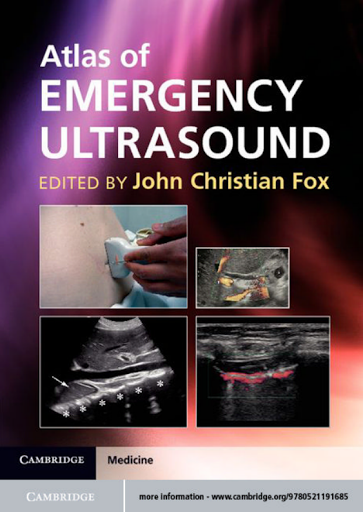
Atlas of Emergency Ultrasound

Complex Cases in Echocardiography

Complex Cases in Echocardiography is a unique text written by a team of top cardiologists at Cedars Sinai Heart Institute. Drawing from their extensive library of echocardiograms, the authors feature 75 cases demonstrating uncommon and puzzling echo findings. Each case begins with a brief clinical presentation, and related images, followed by multiple-choice questions. Detailed answers include patient outcomes and follow-up recommendations. The cases include m-mode, 2-D, 3-D echo, TEE, and Doppler ultrasound. In addition, three fold-out matching quizzes present a particular physical finding or electrocardiogram to be matched with the patient’s corresponding echocardiogram.
With the inclusion of cases seen in emergency situations as well as routine readings in an echo laboratory, this text not only introduces the reader to unusual cases, but also reinforces relevant techniques and applications.. A companion website includes the fully searchable text and features additional cases, along with actual case video clips.
FEATURES:
- Case-based format featuring 75 unusual and bizarre diagnostic dilemmas
- Questions and answers associated with each case
- 3 unique fold-out quizzes
- A companion website with fully searchable text and 45 additional cases
Practical Handbook of Echocardiography: 101 Case Studies 1st Edition

Practical Handbook of Echocardiography: 101 Case Studies Echocardiography is now one of the most commonly used diagnostic imaging tools, yet many clinicians remain unaware of the range of conditions echo can reveal or how echo can be used to help plan therapy. Moreover, it can be quite challenging even for the most seasoned practitioners to spot unusual conditions. Compiled by three echocardiographers with more than 100 years of clinical experience between them, Practical Handbook of Echocardiography uses a case-based approach to explain in detail the full spectrum of echocardiographic modalities and how to optimize their use in the clinical setting. This practical new book: * Covers the full gamut of echocardiographic modalities, including M-mode, 2-D,3-D and Doppler (PW, CW, color flow, tissue and strain), transesophageal (intra-operative and routine) and contrast * Describes cases in both clinical and echocardiographic terms including very interesting cases and the new clinical techniques * Features beautifully reproduced, well-labeled, full color echocardiograms * Includes accompanying DVD with real-time video clips Appropriate for physician echocardiographers and all cardiologists, as well as echocardiographic technicians, Practical Handbook of Echocardiography is the ideal concise guide to using echocardiography to make definitive diagnoses and improve patient outcomes.
Thứ Ba, 18 tháng 8, 2015
Diagnostic Ultrasound – Rumack, 2-Volume Set, 4e

Diagnostic Ultrasound, edited by Carol M. Rumack, Stephanie R. Wilson, J. William Charboneau, and Deborah Levine, presents a greater wealth of authoritative, up-to-the-minute guidance on the ever-expanding applications of this versatile modality than you'll find in any other single source. Preeminent experts help you reap the fullest benefit from the latest techniques for ultrasound imaging of the whole body...image-guided procedures...fetal, obstetric, and pediatric imaging...and more. This completely updated 4th Edition encompasses all of the latest advances, including 3-D and 4-D imaging, fetal imaging, contrast-enhanced ultrasound (CEUS) of the liver and digestive tract, and much more - all captured through an abundance of brand-new images. And now, video clips for virtually every chapter allow you to see the sonographic presentation of various conditions in real time!
- Compare your findings to approximately 5,000 outstanding imaging examples (1,150 in full color).
- Gain valuable diagnostic tips and insights from the most respected experts in the field.
- See the sonographic presentation of various conditions in real time, thanks to video clips accompanying virtually every chapter!
- Master all of the latest US applications, including the newest developments in 3-D and 4-D imaging, fetal imaging, contrast-enhanced ultrasound (CEUS) of the liver and digestive tract, and much more.
- View state-of-the-art examples of all imaging findings with more than 70% new illustrations in the obstetrics section (including correlations with fetal MRI), and more than 20% new images throughout the rest of the contents.
The best-selling reference in Diagnostic Ultrasound, completely revised and updated with new images and expanded use of video clips
Thứ Hai, 17 tháng 8, 2015
Practical Ultrasound: An Illustrated Guide, Second Edition

In the hands of a skilled operator, ultrasound scanning is a simple and easy procedure. However, reaching that level of proficiency can be a long and tedious process. Commended by the British Medical Association, Practical Ultrasound, Second Edition focuses on the scans regularly encountered in a busy ultrasound department and provides everything practitioners need to know to become competent and skilled in scanning.
See What’s New in the Second Edition:
- New chapters on breast, musculoskeletal, and FAST (focused assessment with sonography in trauma) ultrasonography
- Revisions to original chapters incorporating up-to-date techniques and protocols
Beginning with the general principles of ultrasound scanning and a guide to using the ultrasound machine, the book provides step-by-step instructions on how to perform scans supplemented by high-quality images and handy tips.
Organized according to anatomical site, the chapters include a review section on useful anatomy, scan protocol presented step by step, and a section on common pathology. Maintaining the popular format of the previous edition, each chapter contains examples of common and clinically relevant pathologies and notes on the salient features of these conditions.
The authors’ precise approach puts an immense amount of knowledge within easy reach, making it an ideal aid for learning the practicalities of ultrasound.
Thứ Hai, 15 tháng 6, 2015
Ultrasound-Guided Regional Anesthesia and Pain Medicine 2e

Get up-to-date on all of the techniques that are rapidly becoming today’s standard of care with Ultrasound-Guided Regional Anesthesia and Pain Medicine, 2nd Edition. With this extensively revised edition, you’ll see how the increased use of ultrasound for diagnosis and treatment of chronic pain and other medical conditions can transform your patient care. Noted authorities discuss the techniques you need to know for upper and lower extremity blocks, truncal blocks, pain blocks, trauma and critical care, and more.
Key Features:
- Quickly grasp the salient features of each block, including indications, relevant anatomy, the transducer type, needle, local anesthetic, technique, tips, and references for further study.
- Learn from global experts on topics such as recent ultrasound-guided musculoskeletal blocks and the uses of ultrasound in the treatment of shock and trauma.
- Get up to speed on new techniques, thanks to 48 new chapters covering transforaminal cervical injections, cervical facet injections, insertion of peripheral nerve stimulators using ultrasound guidance, genital femoral nerve injections, suprascapular nerve injection, ultrasound guidance to fill implanted pain reservoir, sacroiliac injections, and much more.
- Care for your young patients more effectively with a separate section devoted to pediatric regional anesthesia.
- See exactly how to perform the techniques with high-quality ultrasound images and depictions of relevant anatomy.
Now with the print edition, enjoy the bundled interactive eBook edition, offering tablet, smartphone, or online access to:
- Videos of selected procedures.
- Complete content with enhanced navigation.
- Powerful search tools and smart navigation cross-links that pull results from content in the book, your notes, and even the web.
- Cross-linked pages, references, and more for easy navigation.
- Highlighting tool for easier reference of key content throughout the text.
- Ability to take and share notes with friends and colleagues.
- Quick reference tabbing to save your favorite content for future use.
Thứ Ba, 2 tháng 6, 2015
Ultrasound Teaching Manual: The Basics of Performing and Interpreting Ultrasound Scans

Thứ Bảy, 4 tháng 4, 2015
Endosonography 3rd Ed

From diagnostic to therapeutic procedures, Endosonography, 3rd Edition is an easy-to-access, highly visual guide covering everything you need to effectively perform EUS, interpret your findings, diagnose accurately, and choose the best treatment course. World-renowned endosonographers help beginners apply endosonography in staging cancers, evaluating chronic pancreatitis, and studying bile duct abnormalities and submucosal lesions. Practicing endosonographers can learn cutting-edge techniques for performing therapeutic interventions such as drainage of pancreatic pseudocysts and EUS-guided anti-tumor therapy. Meticulous updates, electronic access to the fully searchable text, videos detailing various methods and procedures-and more-equip you with a complete overview of all aspects of EUS.
"The book is a comprehensive reference book and a real "cookbook" for endosonography."Reviewed by: Professor Abdel Meguid Kassem, Arab Journal of Gastroenterology Date: Jan 2015 "The updated information provided in this book based on experts' knowledge and evidence-based medicine gives an excellent tool for learning EUS and for improving EUS guided therapeutic procedures." Reviewed by: J Gastrointestin Liver Dis Reviewed by: Dec 2014
- Get a clear overview of everything you need to know to establish an endoscopic practice, from what equipment to buy to providing effective cytopathology services.
- Understand the role of EUS with the aid of algorithms that define its place in specific disease states.
- Gain a detailed visual understanding and mastery of how to perform EUSsystematically using illustrations, high-quality endosonography images, and videos.
- Glean all essential, up-to-date information about endosonography including transluminal drainage procedures, contrast-enhanced EUS, and fine-needle aspiration techniques.
- Benefit from the extensive knowledge and experience of world-renowned leadersin endosonography, Drs. Robert H. Hawes, Paul Fockens, and Shyam Varadarajulu.
- Locate information quickly and easily through a consistent chapter structure, with procedures organized by body system.
- Access the full text online at Expert Consult.
Master how to perform EUS systematically using the station-based approach and the latest techniques on FNA and therapeutic interventions using step-by-step procedural videos and high-quality images from leading global authorities.
Thứ Tư, 1 tháng 4, 2015
Interventional Ultrasound - A Practical Guide and Atlas, 1E

The first comprehensive, multi-specialty text on ultrasound guidance in interventional procedures, this book uses the authors extensive clinical experience to provide a full overview of modern interventional ultrasound. For all practitioners, whether new to the procedures or already using them, Interventional Ultrasound offers expert advice and solutions to commonly encountered questions and problems.
Special Features:
- Provides a complete approach to interventional ultrasound, beginning with essential basics on materials, equipment, setup requirements, informed consent issues, microbiologic aspects, and hygiene
- Covers specific, ultrasound-guided diagnostic and therapeutic interventions in the abdomen, thorax, urogenital tract, musculoskeletal system, thyroid and other sites, including indications, selection of materials and biopsy devices, preparation and detailed, hands-on techniques as well as management of complications
- Describes key recent advances, such as the use of ultrasound contrast agents in interventional procedures, adapting ultrasound transducers for endoscopic use in biopsies of the thorax and gastrointestinal tract, performing percutaneous biopsy aspiration and drainage with ultrasound, employing sonography in advanced ablative techniques and more
- Explores such cutting edge topics as symptom-oriented palliative care interventions, applications in critical care medicine and interventions in children
- Highlights, for the first time, the vital role of assisting personnel in interventional ultrasound procedures
Offering easy-to-follow instructions and nearly 600 high-quality illustrations, Interventional Ultrasound takes a practical, cookbook approach ideal for daily use in the hospital or clinic. It is an indispensable reference for interventional radiologists, gastroenterologists, internists, surgeons and other specialists who need to stay up-to-date on the newest technology and applications in this rapidly advancing field.
Chủ Nhật, 28 tháng 12, 2014
Point Of Care Ultrasound 1e

- Access all the facts with focused chapters covering a diverse range of topics, as well as case-based examples that include ultrasound scans and videos.
- Understand the pearls and pitfalls of point-of-care ultrasound through contributions from experts at more than 30 institutions.
- View techniques more clearly than ever before. Illustrations and photos include transducer position, cross-sectional anatomy, ultrasound cross sections, and ultrasound images.
- Expert Consult eBook version included with purchase. This enhanced eBook experience allows you to watch more than 200 ultrasound videos from real-life patients that demonstrate key findings; these videos are complemented by anatomical illustrations and text descriptions to maximize learning. You'll also be able to search all of the text, figures, and references from the book on a variety of devices.
- English | ISBN: 145577569X | 2014 | 456 pages | PDF | 70 MB | 1e
Thứ Bảy, 22 tháng 11, 2014
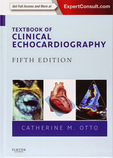
Textbook of Clinical Echocardiography 5th Edition

Textbook of Clinical Echocardiography, 5th Edition enables you to use echocardiography to its fullest potential in your initial diagnosis, decision making, and clinical management of patients with a wide range of heart diseases. World-renowned cardiologist Dr. Catherine M. Otto helps you master what you need to know to obtain the detailed anatomic and physiologic information that can be gained from the full range of echo techniques, from basic to advanced.
- Get straightforward explanations of ultrasound physics, image acquisition, and major techniques and disease categories - all with a practical, problem-based approach.
- Make the most of this versatile, low-cost, low-risk procedure with expert guidance from one of the foremost teachers and writers in the field of echocardiography.
- Know what alternative diagnostic approaches to initiate when echocardiography does not provide a definitive answer.
- Access the entire text online at www.expertconsult.com, as well as echo video recordings that correspond to the still images throughout the book.
- Acquire a solid foundation in the essentials of advanced echocardiography techniques such as contrast echo, 3D echo, myocardial mechanics, and intraoperative transesophageal echocardiography.
- Fully understand the use of echocardiography and its outcomes with key points that identify the must-know elements in every chapter, and state-of-the-art echo images complemented by full-color comparative drawings of heart structures.
- Familiarize yourself with new ASE recommendations for echocardiographic assessment of the right heart and 3D echocardiography, including updated tables of normal measurements.
-
English | ISBN: 1455728578 | 2013 | 548 pages | PDF | 73 MB | 5th
![Download this book here! icon-download[7]](https://blogger.googleusercontent.com/img/b/R29vZ2xl/AVvXsEh2onCUEr3wBLryx3ZQqRbKWea2zNrUfzXUychitFq2711fkmhkCe1A21SM9tZ9PvMJzZCOZxgV9LFCkcfVKVd9iN7R2-7-fGp8o9rymAGKdSaBWQMvp51OsPujKMKYq3LM5cGpIiOwObxp/)


