Latest News

Donate if you like this blog

Hiển thị các bài đăng có nhãn 2007. Hiển thị tất cả bài đăng

Thứ Hai, 27 tháng 4, 2015

The Human Central Nervous System A Synopsis and Atlas 4th Ed

The present edition of The Human Central Nervous System differs considerably from its predecessors. In previous editions, the text was essentially confined to a section dealing with the various functional systems of the brain. This section, which has been rewritten and updated, is now preceded by 15 newly written chapters, which introduce the pictorial material of the gross anatomy, the blood vessels and meninges and the microstructure of its various parts and deal with the development, topography and functional anatomy of the spinal cord, the brain stem and the cerebellum, the diencephalon and the telencephalon. Great pains have been taken to cover the most recent concepts and data. As suggested by the front cover, there is a focus on the evolutionary development of the human brain. Throughout the text numerous correlations with neuropathology and clinical n- rology have been made. After much thought, we decided to replace the full Latin terminology, cherished in all previous editions, with English and Anglicized Latin terms. It has been an emotional farewell from beautiful terms such as decussatio hipposideriformis W- nekinkii and pontes grisei caudatolenticulares. Not only the text, but also the p- torial material has been extended and brought into harmony with the present state of knowledge. More than 230 new illustrations have been added and many others have been revised. The number of macroscopical sections through the brain has been extended considerably. Together, these illustrations now comprise a complete and convenient atlas for interpreting neuroimaging studies.

  • English | ISBN: 3540346848 | 2007 | 966 pages | PDF | 88 MB | 4th

    Download

  • Thứ Năm, 9 tháng 4, 2015

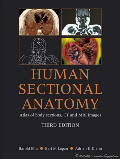
    Human Sectional Anatomy Atlas of Body Sections, CT and MRI Images, Third Edition

    First published in 1991, Human Sectional Anatomy set new standards for the quality of cadaver sections and accompanying radiological images. Now in its third edition, this unsurpassed quality remains and is further enhanced by some useful new material.
    As with the previous editions, the superb full-colour cadaver sections are compared with CT and MRI images, with accompanying, labelled line diagrams. Many of the radiological images have been replaced with new examples, taken on the most up-to date equipment to ensure excellent visualisation of the anatomy. Completely new page spreads have been added to improve the book's coverage, including images taken using multidetector CT technology, and some beautiful 3D volume rendered CT images. The photographic material is enhanced by useful notes, extended for the third edition, with details of important anatomical and radiological features.

  • English | ISBN: 0340912227 | 2007 | 287 pages | PDF | 33 MB | 3rd

    Download

  • Thứ Bảy, 14 tháng 3, 2015

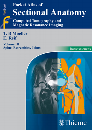
    Pocket Atlas of Sectional Anatomy - Computed Tomography and Magnetic Resonance Imaging, Volume III

    Renowned for its superb illustrations and highly practical information, the third edition of this classic reference reflects the very latest in state-of-the-art imaging technology. Together with Volumes 1 and 2, this compact and portable book provides a highly specialized navigational tool for clinicians seeking to master the ability to recognize anatomical structures and accurately interpret CT and MR images.Highlights of Volume 3:

  • New CT and MR images of the highest quality
  • Didactic organization using two-page units, with radiographs on one page and full-color illustrations on the next
  • Concise, easy-to-read labeling on all figures
  • Color-coded, schematic diagrams that indicate the level of each section
  • Sectional enlargements for detailed classification of the anatomical structureComprehensive, compact, and portable, this popular book is ideal for use in both the classroom and clinical setting.
    • English | ISBN: 158890475X | 2007 | 329 pages | PDF | 110 MB | 3rd


    Download

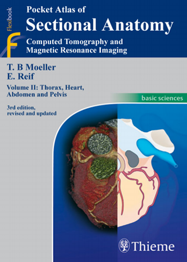
  • Pocket Atlas of Sectional Anatomy - Computed Tomography and Magnetic Resonance Imaging, Volume II

    Known to radiologists around the world for its superior illustrations and practical features, Pocket Atlas of Sectional Anatomy reflects the very latest in imaging technology. It provides in-depth coverage of the thorax, heart, abdomen, and pelvis in a user-friendly format. In the clinic, this compact book acts as the perfect navigational tool for radiologists and technicians with CT and MRI.

    Highlights of Volume II: Thorax, Heart, Abdomen, and Pelvis

  • Practical two-page units comprised of radiographs on one page and full-color schematics on the next
  • More than 440 clearly labeled illustrations
  • Many new CT and MR images of the highest quality accompany stunning, full-color drawings, now enhanced with cardiac imaging
  • Consistent color coding makes it easy to identify individual structures across several contiguous sections

    Volume II: Thorax, Heart, Abdomen, and Pelvis and its companion books -- Volume I: Head and Neck and Volume III: Spine, Extremities, Joints -- comprise a must-have resource for radiologists of all levels.

    • English | ISBN: 158890475X | 2007 | 257 pages | PDF | 69 MB | 3rd


    Download

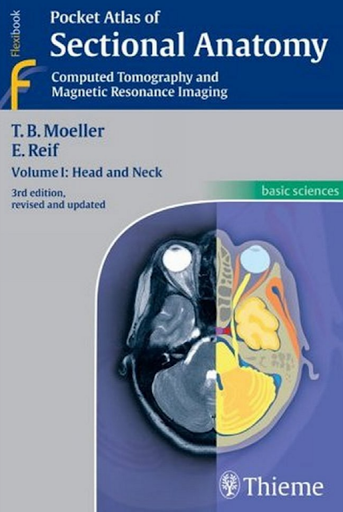
  • Pocket Atlas of Sectional Anatomy - Computed Tomography and Magnetic Resonance Imaging, Volume I

    Renowned for its superb illustrations and highly practical information, the third edition of this classic reference reflects the very latest in state-of-the-art imaging technology. Together with Volumes 2 and 3, this compact and portable book provides a highly specialized navigational tool for clinicians seeking to master the ability to recognize anatomical structures and accurately interpret CT and MR images.

    Features:

    • New CT and MR images of the highest quality
    • Didactic organization using two-page units, with radiographs on one page and full-color illustrations on the next
    • Concise, easy-to-read labeling on all figures
    • Color-coded, schematic diagrams that indicate the level of each section
    • Sectional enlargements for detailed classification of the anatomical structure

    Comprehensive, compact, and portable, this book is ideal for use in both the classroom and clinical setting.

    • English | ISBN: 158890475X | 2007 | 272 pages | PDF | 26 MB | 3rd


    Download

    Thứ Sáu, 20 tháng 2, 2015

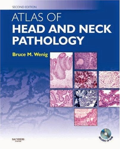
    Atlas of Head and Neck Pathology 2nd Ed

    Effectively and efficiently diagnose the full range of neoplastic and non-neoplastic diseases of the head and neck with this peerless source of pathology guidance! 2,240 phenomenal illustrations depict the histologic, immunohistochemical, cytologic, and diagnostic imaging appearance of every type of head and neck pathology, including brand-new coverage of thyroid and parathyroid disorders. You'll also find completely new guidance on identifying intraoperative specimens, applying the latest molecular biological and cytogenetic techniques, and much more. A consistent, practical organization and succinct, bulleted format make the reliable answers you need easy to find.

    • Review each disease's clinical features, pathology, differential diagnosis, treatment, and prognosis quickly thanks to a concise, user-friendly, bulleted format.
    • Consider cytopathology, special stains, and cytogenetics where appropriate.
    • Compare different disease types at a glance with numerous tables, charts, and helpful lists.
    • Identify specimens confidently by comparing them to 2,240 illustrations-most in full color-capturing the histologic, immunohistochemical, cytologic, and diagnostic imaging appearance of every form of head and neck pathology.
    • Diagnose a broader range of head and neck diseases with new coverage of thyroid and parathyroid disorders.
    • Provide effective intraoperative consultations with guidance on identifying frozen mucosal, salivary gland, thyroid, and parathyroid specimens that's hard to find in other texts.
    • Apply the latest WHO classification guidelines and AJCC staging criteria for head and neck neoplasms.
    • Perform the newest molecular biologic and cytogenetic techniques.
    • English | ISBN: 0721697887 | 2007 | 1042 pages | PDF | 349 MB | 2nd


    Download

    Thứ Tư, 30 tháng 7, 2014

    Emergency Radiology Case studies

    Effectively and confidently interpret even the most challenging radiographic study

    A Doody's Core Title!

    "...should be a part of every emergency medicine resident's personal library. In addition to residents, I would highly recommend this book to medical students, midlevel providers and any other physician who is interested in improving their ability to interpret radiographic studies necessary to diagnose common emergency medicine patient complaints."--Annals of Emergency Medicine

    4 STAR DOODY'S REVIEW!
    "The purpose is to help improve the reader's skills in ordering and interpreting radiographs. The focus is on conventional radiographs, as well as noncontrast head CT. For emergency physicians this is a vital skill, which can greatly aid in making difficult diagnoses. The book is well written and thorough in addressing how to read radiographs, as well as covering easy to miss findings. The numerous pictures and radiographs are invaluable in demonstrating the author's teaching points and in engaging the reader in the clinical cases....This well written book will be extremely useful for practicing emergency physicians. The clinical cases are interesting and help challenge the reader to improve their skills at evaluating radiographs more thoroughly."--Doody's Review Service

    Emergency Radiology: Case Studies is a one-of-a-kind text specifically designed to help you fine-tune your emergency radiographic interpretation and problem-solving skills. Illustrated with hundreds of high-resolution images, this reference covers the full range of clinical problems in which radiographic studies play a key role. Dr. David Schwartz, a leading educator, takes you step-by-step through the radiographic analysis of medical, surgical, and traumatic disorders, giving you an unparalleled review of the use and interpretation of radiographic studies in emergency diagnosis.

    Features

    • 55 cases studies that highlight challenging areas in emergency diagnosis, including imaging studies with subtle, equivocal, or potentially misleading findings
    • Detailed coverage of the broad spectrum of disorders for which radiographs are utilized in emergency practice
    • Coverage of chest and abdominal radiology, the extremities, cervical spine and facial radiology, and head CT
    • Cohesive template for each chapter, beginning with a case presentation, followed by a comprehensive discussion of the disorder under consideration
    • Sections begin with an overview of the pertinent radiographic technique, anatomy, and method of radiographic interpretation
    • Diagnosis-accelerating radiographs, ultrasound images, CT scans, and MR images
    • Invaluable “pearls and pitfalls” of radiographic interpretation

    Pages: 555

    Size: 39 Mb


    Download

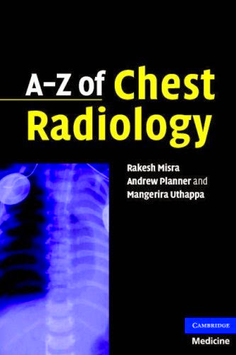
    A - Z of Chest Radiology

    A-Z of Chest Radiology provides a comprehensive, concise, easily accessible radiological guide to the imaging of acute and chronic chest conditions. Organised in A-Z format by disorder, each entry gives easy access to the key clinical features of a disorder. An introductory chapter guides the reader in how to review chest X-ray's accurately. This is followed by a detailed discussion of over 60 chest disorders, listing characteristics, clinical features, radiological features and management. Each disorder is highly illustrated to aid diagnosis; the management advice is concise and practical. A-Z of Chest Radiology is an invaluable quick pocket reference for the busy clinician as well as an aide memoir for revision in higher exams in both medicine and radiology.
    Pages: 224
    Size: 7 Mb

    Translate

    Total Pageviews